New ROM exhibit digitally unravels inner remains of Egyptian mummies
[ad_1]
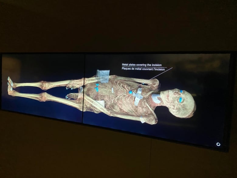
If you’ve ever wondered what exactly lies beneath the linen wrappings of ancient mummies, a new exhibit at the Royal Ontario Museum uses modern technology to peel through the layers and reveal the remains — all without unravelling their bandages.
“In the 19th century, people were studying the mummies by unwrapping them. In the 20th century, it was X-ray. In the 21st century, we’ve got fancy CT scans — three-dimensional ones that in this case, leads to some interesting results,” said Krzysztof Grzymski, the ROM’s senior curator of Egypt and Nubia.
Egyptian Mummies: Ancient Lives, New Discoveries features six mummified individuals who range from a young child to middle-age adults. It showcases their life stories and how they lived over 3,000 years ago along the Nile River.
The exhibit, which opens this weekend and is on loan from the British Museum, runs until March 2021.
Its research team was the first to use advanced CT scanners and software on the mummies and see high-definition images of their remains, according to Grzymski.
“With this technique we don’t need to unwrap the mummy,” he said.
From the images, researchers were able to identify key information like the individual’s sex, age and health at the time of death.
“The exhibition is not only about the mummies and the dead. We also try to reconstruct their life and their daily routine,” Grzymski said, noting there are showcases of instruments, jars, and children’s toys from the time.
CT scans revealed dental problems
The three-dimensional images show amulets — used as good luck charms — placed in various parts of the mummies’ bodies. Researchers were able to recreate some of the objects using 3D printing.
CT scans revealed clearly that four of the six mummies had dental problems, which Grzymski attributes to an unhealthy diet of sweets like honey and figs and small fragments of rock and sand that would get into their food.
“We can see the abscesses in their teeth. It wore the teeth down and they were suffering,” he said.
Mummification, an ancient practice done to preserve the body for the afterlife, was used for people of different classes. According to Grzymski, the coffins and wrappings of the six mummies reveal they were from lower and upper middle classes.

He said the first mummy on display, Nestawedjat, was a married woman from an established middle-class family based on the fact she was placed in three coffins — the innermost one being ornately decorated and shaped like a human figure.
Based on inscriptions, it was first believed the coffin belonged to a female, but a subsequent X-ray made researchers believe it held a male’s body based on its body structure.
It wasn’t until the British researchers used CT scans that it was determined the body was indeed of a female. Her body had been carefully mummified and fingerprints were left by the embalmers who used resin for preservations.

Grzymski also noted that the first CT scans performed on mummies was done in Toronto in 1974 by doctors and researchers at SickKids.
He imagines one day researchers will be able to discover more about the genetics of mummies.
“We are fortunate to have soft tissues preserved to those early humans,” he said. “Perhaps with the use of advanced studies of genetic material, we can learn something more about those people, about the relationship between them and other cultures and other civilizations.”
ROM taking COVID-19 precautions
Ancient Mummies is a ticketed event that limits the number of visitors in attendance. The museum is asking both staff and visitors to be masked.

“We were able to design the exhibition itself in such a way that there is plenty of distance between the cases,” Grzymski said.
“In times of COVID-19, it’s really important that we can lay out the exhibition in such a way that it’s safe for visitors.”
[ad_2]
SOURCE NEWS
